UPMC/University of Pittsburgh Schools of the Health Sciences
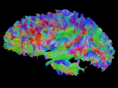
Above: High definition fiber-tracking of a million brain fibers. Click the photo to download it in high resolution. Photo Credit: Walt Schneider Laboratory
A powerful new imaging technique called High Definition Fiber Tracking (HDFT) will allow doctors to clearly see for the first time neural connections broken by traumatic brain injury (TBI) and other disorders, much like X-rays show a fractured bone, according to researchers from the University of Pittsburgh in a report published online today in the Journal of Neurosurgery. HDFT could provide an objective way of identifying brain injury, predicting outcome and planning rehabilitation.
The study was funded by the Defense Advanced Research Projects Agency.
Downloadable Images
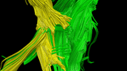
High definition fiber tracking reveals loss of fibers, or connections, on the injured right side (yellow) and the intact, undamaged left side (green). The patient was injured in an ATV accident and lost function in his left leg, arm and hand”.
Press Releases


